Blood–Brain Barrier (BBB) Model: This model has been used for determining the permeability of medicines and advanced therapies (ATMP) under development that, after being approved, will be used for treating neurological disease. Current BBB models are not realistic enough.
Scaffold for Bone Regeneration: This is used for personalised bone regeneration treatment that takes the specific area being treated into account. Current models are not homogeneous and do not reproduce that complexity.
Two different applications of additive manufacturing in the biomedical sector have therefore been consolidated in this project with a single manufacturing methodology.
Scaffolds for Bone Regeneration
The structure of bone defects has been reconstructed using micro-computed tomography (μCT) images that could not be expected to regenerate satisfactorily by natural means because of their dimensions: critical-size bone defect. The resolution of the structure is coarse in comparison with the case, but its volume may be greater (several cm3). The material chosen is a bioabsorbable polymer (polycaprolactone, PCL). The scaffold is then processed by adding osteogenic proteins (morphogenetic bone protein rhBMP-2) and mesenchymal stem cells.
Implants with a 3D structure quite faithful to the bone being regenerated with good connectivity and porosity have been obtained (Figures 1 A, 1B). Additionally, it has been possible to print on 3D scaffolds with a PCL membrane which will make it possible to add different treatments (cells, morphogens) in a way that mimics the physiological response of tissue damage (Figure 1 C-1 F). Printing different scaffolds to meet the demand for implants in animal trials carried out at the CUN was continued in the second half of 2019.
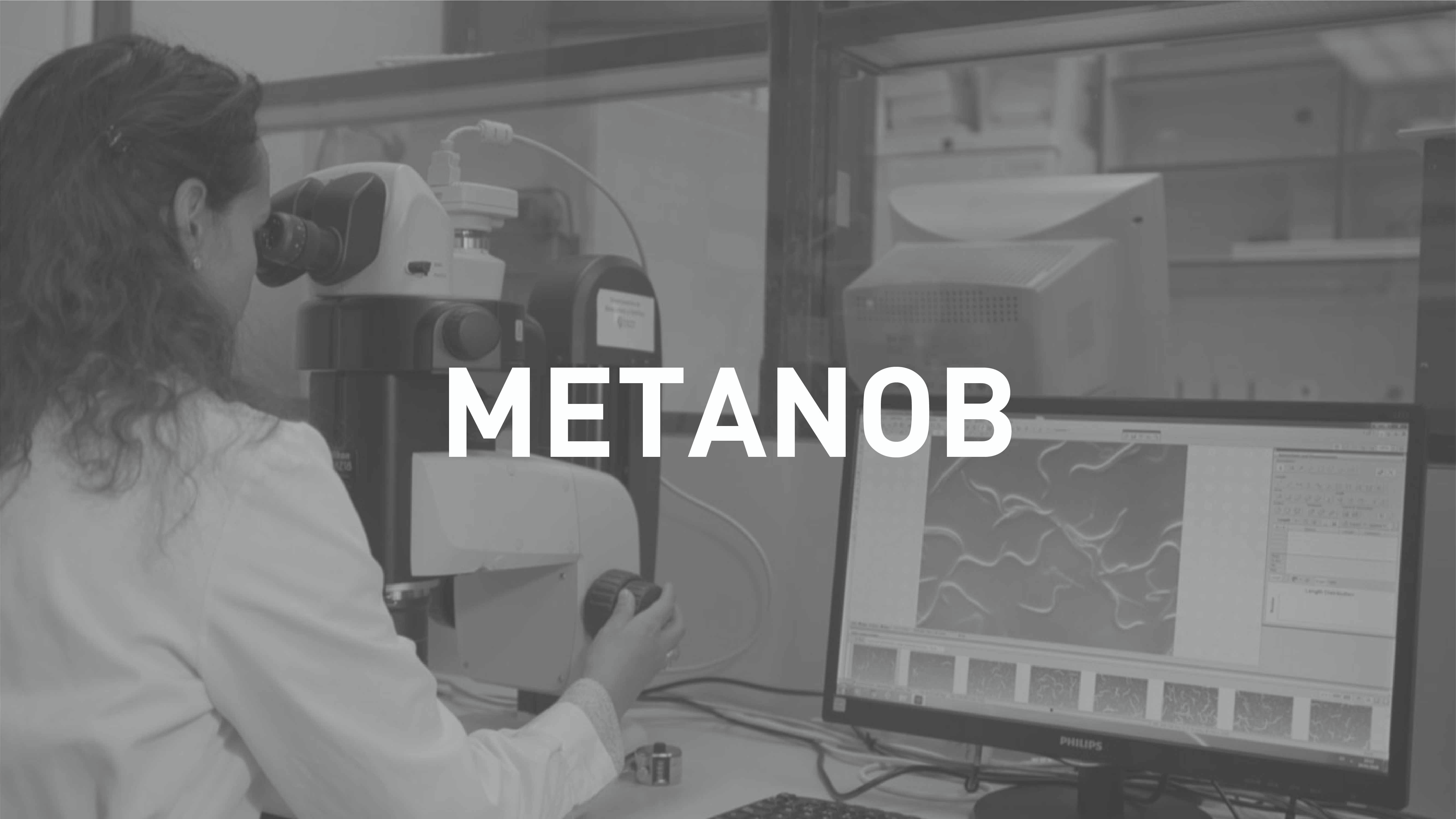
Blood-Brain Barrier Models
A multi-scale model of the three-dimensional barrier has been created using confocal microscopy images of real tissue. Manufacturing resolutions up to 50m and printing volumes of several mm3 have been obtained. These devices are used for in vitro research of medicines that will be used for treating neurodegenerative diseases. The model is used for three-dimensional growth of endothelial cells, pericytes and astrocytes, which are specific sub-populations that simulate the barrier with greater fidelity.
Work has been done on optimising a polyethylene glycol diacrylate (PEGDA) formulation suitable for use in the stereolithography device for Peopoly Moai 3D printing.
The effect of the following parameters on a specific 1.12 gr/ml PEGDA solution concentration has been analysed comparing the results obtained with the design obtained with a reference resin from the device itself: the photoinitiator concentration, absorbent concentration and the strength of the device’s laser.
The central area with the narrowest microchannels is in turn the area where the cellular adhesion occurs asnd the bilayer that makes up the artificial BBB. The adhesion of the epithelial cells is promoted in the inner area, while the pericytes and astrocytes adhere on the outside. The device finishes with a standard microfluidic connection (1/32″ tubing wit C100 connectors).
An initial formulation with PEGDA and a photoinitiator were prepared, in this case 2,2-Dimethoxy-2-phenylacetophenone (DMPA) was used and films were prepared to study cellular adherence on this kind of material.
It is known a priori that PEGDA by itself does not have good cellular adhesion, so it is necessary to superficially functionalize the films with biomolecules that promote cellular adhesion.
The films were produced by Naitec and sent to Navarrabiomed, where they were functionalized with laminins and the cell cultures were done. The cellular adherence was analysed a posteriori with scanning electronic microscopy done by Naitec.